
Best Ultrasound Sonography Centre In Navi Mumbai
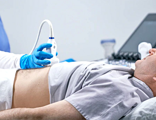
Dr.Vivek's Diagnostic Center was established in July 2005 with the aim of providing cost-effective medical services to the average person with a professional approach. Ultrasound scans create an image of the internal structures of the human body using high frequency sound waves. Ultrasound is widely used by doctors to examine the developing fetus (unborn child), abdominal and pelvic organs, muscles and tendons, heart and human blood vessels. Ultrasound is also known as a sonogram or (when viewing the heart) EchocardiogramHigh-frequency sound waves are sent by the ultrasound machine into the internal structures of the body being examined. The echoes or reflected sounds are collected to form an image that can be viewed on a monitor. The sound waves are sent and received by a small portable transducer. Because of the high frequency of the sound, the human ear cannot hear it, which is why it is called an ultrasound scan. Ultrasound scans are usually non-invasive (performed outside the body). However, some scans are done using a special probe inserted into your vagina (for some obstetric or pelvic exams), rectum (for some prostate exams) or esophagus (for some heart exams).Sometimes doctor use ultrasound to monitor and guide invasive procedures such as breast or thyroid biopsies.If You are Loooking for the Top sonography Centre in Mumbai Then Nexsus Diagnostic Centre in Navi Mumbai is the Best Ultrasound Sonography Centre In Navi Mumbai
Illnesses and ultrasound scans
Some ultrasounds require special preparation, such as abstaining from food for several hours before an upper abdominal scan.
Some pelvic exams require that you have a full bladder before the scan. If you need to do anything out of the ordinary before the scan, consult our doctor or ultrasound service.
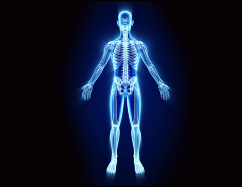
Full Body Sonography
Ultrasound imaging utilizes sound waves to deliver photos of within the body. It is used to examine a baby in pregnant women and the brain and hips in infants, as well as to help diagnose infections, pain, and swelling in the body's internal organs.
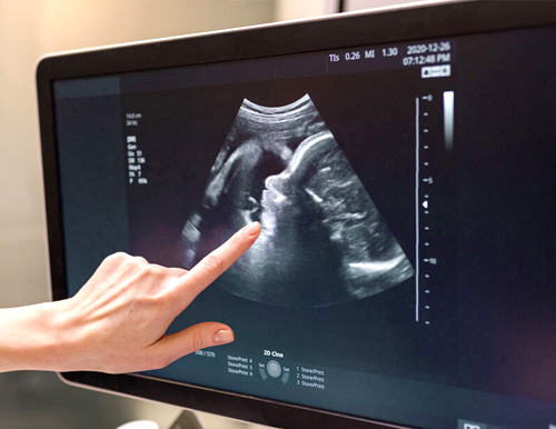
Q1)What exactly is a 3D ultrasound?
Ultrasound, and more recently, complex 3D ultrasound scans, are now one of the best technologies for viewing the insides of the body.These scans produce stunning, high-resolution, three-dimensional images.Dr Vivek Ukirde he has 20 years of experience in the Field of radiologist and interventional radiologist.He has authored couple of book in interventional radiology in Superficial Femoral Artery.
Q2)Advantages of 3D Ultrasound For Newborns
Since 3D ultrasound images are much clearer and sharper, they can help the physician detect any problems in the baby's growth.
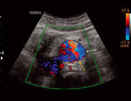
Color Doppler Test
A color Doppler test is a diagnostic procedure that uses sound waves to create a picture. It reports on the speed, velocity and direction of blood flow. This test will be used by your doctors to look for blockages and clots in your blood vessels. This is not possible because conventional ultrasonography does not show blood flow. No dye is injected into your body during a color Doppler examination. As a result, this is a safe and convenient test. The color doppler test uses the Doppler effect. This technique creates images of blood flow by measuring sound waves reflected from moving objects, such as Red Blood Cells
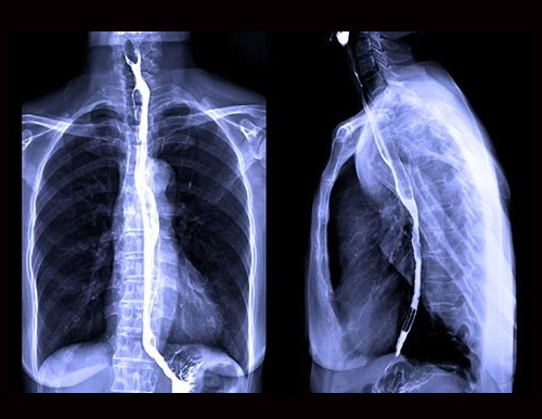
Barium Procedures
The Radiologist will ask you to swallow a thick, chalky barium drink. The barium is typically flavored, but it may not be very good. After you swallow the barium, the radiologist will take single images, a series of X-rays, or fluoroscopy to observe how it moves through your mouth and throat.
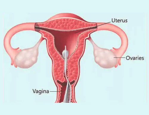
HSG
Hysterosalpingography, also known as uterosalpingography, is a radiologic procedure to investigate the shape of the uterine cavity and the shape and patency of the Fallopian tubes. It is a special x-ray using dye to look at the womb and Fallopian tubes.
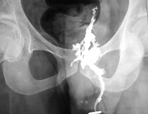
Fistulogram
A particular x-ray procedure is called a fistulogram. It utilizes contrast (x-beam color) to take a gander at the blood stream in your fistula or join (dialysis access). This methodology can verify whether it is hindered or on the other hand assuming that there is any restricting (stenosis)
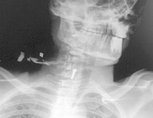
Sinogram
A sinus examination is done with a sinogram. A sinus resembles a fistula. However, rather than connecting two organs, it resembles a closed tube at one end. The special dye that is injected into the fistula or sinus is used in both of these tests. The dye makes it possible for the X-ray to show the sinus or fistula.
Micturating Cystourethrography
A scan called a micturating cystourethrogram (MCUG) depicts your child's bladder function. It is utilized to determine the cause of your child's possible urinary tract infections. Additionally, it is used to detect any urinary system issues in your child.Want to know more about the Best ultrasound sonography centre in Navi Mumbai.Visit our Website for more information

IVP - Intra Venous Pyelogram
IVP An IVP takes pictures of the urinary tract (kidneys, ureters, and bladder) using X-rays and a special dye that is injected into your veins. An X-ray machine is used to take pictures after the dye is injected. The color will drop of your body in your pee. You won't see it as it is dismal.
The Advantages of using an IVP to examine your kidneys, ureters, and bladder in greater detail during diagnosis
